PlusmanLLC

April 2023

Mathematical Medicine Vol. 3

Interview with a Leading Expert in Medical AI

Becoming More Positive Toward AI Benefits Both Radiology and Patients

Dr. Sadayuki Murayama

Professor Emeritus, University of the Ryukyus
Former Director, Japan Radiological Society
Special Member, Japan Lung Cancer Society
Secretary, Chest Radiology Study Group
Former Chair, Respiratory Function Imaging Study Group

AI Is Extremely Useful Even for Top Experts

Dr. Sadayuki Murayama, a former board director of the Japan Radiological Society and Professor Emeritus at the University of the Ryukyus, has achieved remarkable accomplishments in the field of chest radiology. Why did he ultimately choose Plusman’s AI after trying various systems? How has it helped with interpretation? We spoke with him about clinical communication using AI, ideal users, chest X-ray AI, and finally, his message to young doctors.

Dr. Sadayuki Murayama ,Professor Emeritus, University of the Ryukyus

Accuracy and Lymph Node Visualization Are Effective in Interpretation

Plusman: Dr. Murayama, you have made many outstanding contributions in radiologic diagnosis, particularly in chest imaging. We imagine that many AI vendors have approached you, and you’ve had opportunities to try various AI systems. Ultimately, you chose Plusman’s AI, Plus.Lung.Nodule (Dr. Murayama gave the nickname PLN to Plus.Lung.Nodule. hereafter “PLN”). What were the reasons behind that decision?

※Note: Regulated medical device: System for providing general image diagnostics workstation / Product name: Plus.Lung.Nodule / Certification number: 301AGBZX00004000 / Manufacturer/distributor: Plusman LLC

Dr. Murayama: I had seen products from other companies as well, but I had a willingness from the start to adopt Plusman’s product if it seemed good, and I was glad I was able to use it during the trial period. After comparing it with other products and verifying its accuracy, I found it extremely effective for highlighting regions of interest for lung nodules and aiding interpretation. Also, PLN overlays circular markers directly on the original CT images, which, once you get used to, makes it very easy to read. That strongly influenced my decision to adopt it. What made it even more appealing was that, unlike other products, PLN also visualizes lymph node regions of interest.

Being a startup, I had the impression that Plusman would be agile. One of the advantages of working with a startup is that they sincerely address the feedback I provide.

Plusman: You mentioned that lymph node visualization is one of the differentiators. How do you utilize that feature in clinical practice?


Dr. Murayama: Chest CTs performed with contrast are often used for detailed lung cancer evaluation or for mediastinal tumor assessment, but usually we interpret lung nodules and lymph nodes using non-contrast CTs. What I was most concerned about was that when there is hilar lymphadenopathy on the right side, it can be hard to interpret, and I sometimes ended up concluding “no lymph node enlargement,” which left me feeling uneasy. That’s because the hilar region is continuous with the pulmonary artery, making it difficult to distinguish between the two. Plusman had a demo on their website comparing contrast-enhanced and non-contrast CTs, and the area I was concerned about was clearly visualized even on plain CT. That was very intriguing, and although I had some doubts about whether it would be practically usable, it piqued my interest.

Even Specialists Should Use PLN, Not Just Generalists

Plusman: Having actually used PLN, what kind of doctors or medical facilities do you think it is suited for?

Dr. Murayama: I often hear about research or actual clinical usage of AI like this, but it seems that chest imaging specialists aren’t really using it much. First of all, I think it’s highly useful for people who aren’t familiar with interpreting chest CTs. Traditionally, CAD (Computer-Aided Detection) hasn’t been seen as very helpful for specialists, but it’s said to be extremely useful for non-specialist readers or beginners. In fact, when we started using PLN, just like the literature says, nodules located next to central vessels can be confusing because they look like vascular cross-sections. There was a case during the early stages of PLN implementation where a nodule adjacent to a vessel went undetected when comparing with previous images. I’ve used PLN for about six months now, and I’ve encountered cases where I might have missed something if not for PLN. This two-month pre-study period gave me a good opportunity to realize how easy it is to overlook nodules near vessels. There were actual cases where I found missed nodules upon review, and others where I suspected I might have missed something—so it was very educational. PLN also detects many small nodules. I now realize that in the past, I may have simply ignored extremely tiny nodules.

 As for who should use PLN? Well, even specialists can miss things. PLN displays even small nodules consistently, so anyone involved in image interpretation should use it. With PLN, interpretation becomes more reliable.

Plusman: There’s a common narrative that AI is mainly for younger or non-specialist physicians, and that specialists don’t need it. But based on what you’ve seen, it seems PLN can be valuable even for top-tier physicians?

Dr. Murayama: Yes, I truly believe PLN is extremely helpful even for top-level doctors. 

Thanks to PLN, I No Longer Hesitate About Tiny Nodules

A Paradigm Shift

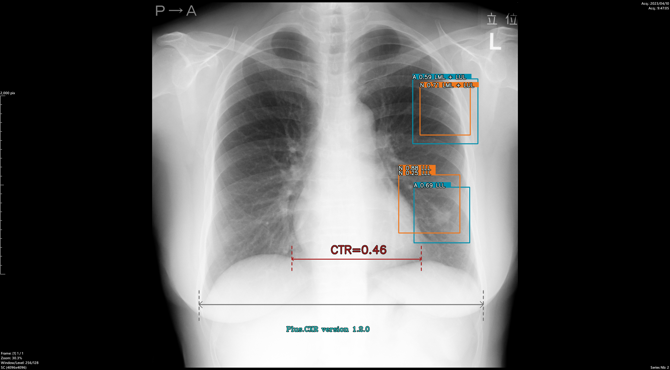
Dr. Murayama: In terms of interpretation time, compared to before, I now spend a bit more time looking at the images carefully. That’s because in the past, I didn’t consciously pay attention to 1–2 mm nodules—I may have thought of them all as parts of blood vessels.But after introducing PLN, I realized that almost everyone has nodules around 2 mm.(※)

Years ago, Dr. Swensen published a paper reporting that about 70% of people had nodules in chest CT screenings. I assumed that was a characteristic of American patients and that fewer Japanese people had nodules. However, after using PLN, I found that most people had nodules ≥2 mm, confirming that what Dr. Swensen said was indeed correct. Now I think it’s natural to assume that almost every patient has nodules of at least 2 mm.

There are intrapulmonary lymph nodes near the pleura, and it turns out such small nodules exist in most people. I struggled at first with how to interpret them, but I now write in reports: “Several small nodules are observed, but they are not considered clinically significant.” For round nodules of ≥3 mm or nodules of ≥5 mm, I pay close attention. Nodules under 3 mm are rarely malignant, but for ≥5 mm, the risk increases, and I make sure to write a detailed report since they could represent early-stage cancer.

So yes, I now understand that there are many even smaller nodules—but I didn’t realize that before using PLN. It truly triggered a paradigm shift in my thinking.

※ Lung Cancer Screening with CT: Mayo Clinic Experience, Stephen J. Swesen, et al., Radiology 2023.3226.3, 756-761

Plusman: For nodules around 2 mm, is the only option to follow them up over time?

Dr. Murayama: That’s right. Literature shows that the likelihood of malignancy in nodules under 5 mm is extremely low, so annual screening is sufficient. There’s no need for short-interval follow-ups. Discovering so many 2 mm nodules thanks to PLN has solidified that belief for me. In the past, even if I found a 3 mm nodule, I would write, “Please follow up.” But in retrospect, that may have been a randomly discovered nodule, and there may have been others I didn’t even see. There are many diagnostic thresholds—like 5 mm or 6 mm—and those are correct. I’ve re-recognized that even if a small nodule appears potentially tumorous, follow-up is rarely necessary unless it’s larger than 5 mm.

Sharing PLN Results with Pulmonologists and Thoracic Surgeons

Plusman: At Urasoe General Hospital, do clinical departments also view the AI results?

Dr. Murayama: PLN results aren’t openly shared with all clinical departments. However, I do attach images showing PLN-detected nodules in the reports I send to pulmonologists and thoracic surgeons. So they know that we’re using PLN and that it marked certain regions of interest. We’re still considering whether to make the terminal accessible to respiratory physicians. Because the viewer shows both nodules and lymph nodes, it might make the interpretation feel cluttered and could be disliked (depending on the viewer, nodules and lymph nodes may be displayed together or separately). Until there’s an explicit request from clinicians, I’m holding off.

Since pulmonologists and thoracic surgeons deal with lung cancer in their day-to-day clinical work, I think they should see the AI output—but maybe not other specialists. While we would need to explain the AI results to each department, I don’t think it’s necessary to go that far just yet. Still, I subtly indicate that we are using it.

Plusman: After sending AI-marked images to respiratory physicians, did you receive any feedback?

Dr. Murayama: One pulmonologist said, “You’re using AI now, right? It’s effective.” A few others probably heard my recent talk about it.

Training Is Needed to Use AI Effectively

Plusman: Let’s say in the future, clinical departments request to use the AI-enabled viewer. What challenges might arise if non-radiologists use diagnostic imaging AI?

Dr. Murayama: As I mentioned earlier, PLN displays nodules that are 2 mm or larger, so the number of detected findings can be quite high. (Note: With PLN, users can customize detection settings such as the threshold for minimum nodule size or the number of nodules to display.) In addition to true nodules, things like curved blood vessels or localized atelectasis under the pleura can also be shown. Those unfamiliar with imaging may be confused—“What is this supposed to be?” That uncertainty can be burdensome, especially for physicians who can’t distinguish whether something is a nodule or not. They may struggle with whether they should mention it as a “nodule” or not. So if AI is going to be widely used, those who use it should first attend a lecture or receive some form of structured training. For example, the CT Screening Society runs a summer seminar that teaches how to identify nodules. Something similar should be done for AI-based interpretation. Just telling beginners, “Use this tool,” will leave them confused—it needs to be taught in seminars or study groups. 

That’s why AI shouldn’t just be sold and left alone. The lung contains many small nodules, and interpreting them can be difficult. A list of false positives is essential—and perhaps even more important than the detection results themselves.

You could reduce the number of displayed findings by increasing the detection threshold to, say, 5 mm instead of 2 mm—but then you would lower the value of AI. To make AI usable by others, the key is how to handle false positives. If users don’t understand which nodules can be safely ignored, it will be overwhelming.On the other hand, if you lower sensitivity, you risk missing true nodules. Personally, I think the current settings are just fine—but I’m not sure how others feel.

Plusman: From a diffusion standpoint—not just in research, but for everyday users—what do you think is necessary for AI to become widely used in clinical settings?

Dr. Murayama: Letting people try it is the most important step. There was a modality in the past that rapidly spread because hospitals were allowed to try it out. Vendors shared both the good and bad feedback with other hospitals, and improvements happened quickly. So I think it’s essential to ask questions and gather feedback from hospitals using AI. Understanding the pain points during real-world usage will be key to adoption.

Growing Diagnostic Value in Interstitial Pneumonia and MRI Diffusion-Weighted Imaging

Why Qualitative Diagnosis Is Still Hard for AI

Plusman: One possible future direction for AI is not just marking areas of interest, but actually calculating diagnostic metrics—like lung cancer volume or VDT (volume doubling time )—and displaying information useful for treatment decisions. What are your thoughts on that?

Dr. Murayama: That would be the next step. Determining whether a pulmonary nodule is benign or malignant falls under qualitative diagnosis. While qualitative diagnosis is often a research topic at academic conferences, I’m not sure how much general physicians will engage in that level of analysis.For example, if there’s a nodule larger than 1 cm, doctors may ask, “Is this cancer or not?” But in routine CT screening, I don’t think that kind of qualitative judgment is always necessary. In the end, it comes down to one of four decisions: ignore it, monitor it closely, biopsy it, or surgically remove it. So I think it’s okay for qualitative diagnosis to be left to the next step. That said, how you judge small nodules is crucial. 

I think current AI isn’t capable of making that kind of nuanced judgment. AI doesn’t reason its way to findings—it just detects features. So for the next step, human input is still necessary. As for using VDT to assess whether a nodule is malignant, I’m looking forward to seeing how that feature works. I think it’ll be useful. But that kind of functionality will likely be used among specialists. For general practitioners or screening centers using PLN, this step may not yet be relevant.

Chest X-ray AI That Excels at Clavicle-Overlapping Nodules

After an upgrade to Plus.CXR(※), Dr. Murayama provided test data for evaluation. The results were analyzed and reviewed.

※Note: Regulated medical device: System for providing general image diagnostics workstation / Product name: Plus.Lung.Nodule / Certification number: 301AGBZX00004000 / Manufacturer/distributor: Plusman LLC

A case of solid lung cancer in the left lower lobe and a part-solid nodule in the outermost part

Dr. Murayama: It’s gotten significantly better! Especially the ability to mark areas around the clavicle—a typical blind spot—is a great improvement. I’d like to summarize this and present it at a conference or study group.

The Ultimate Goal Is to Save Patients—That’s Why We Must Elevate the Level of Medicine

Plusman: Diagnostic value is a function of available treatments. Even if diagnosis is accurate and early, it loses its value if no effective treatment exists. On the flip side, when treatment options improve, precise diagnosis becomes more valuable. With that in mind, are there any diseases or areas where you feel diagnosis has become particularly valuable compared to the past?

Dr. Murayama: In the case of cancer, when various drugs with gene-related antitumor effects and treatments for diseases that previously had no treatments emerge, diagnostic imaging must also evolve accordingly. For example, in the case of chest cancer, antifibrotic drugs are available for interstitial pneumonia. It is necessary to properly diagnose usual interstitial pneumonia (UIP). Until now, it was a disease with no treatment other than lung transplantation. So we looked for evidence of interstitial pneumonia caused by causes other than UIP. If there is such evidence, it will be eligible for steroid treatment, that is, treatment. However, nowadays, if we proactively diagnose UIP, active interstitial pneumonia, we can use antifibrotic drugs, although they are expensive.

MRI will also play a growing role in qualitative imaging. Techniques like diffusion-weighted imaging have been paradigm-shifting. Until now, AI mostly focused on marking areas of interest, but modern imaging needs to support qualitative diagnosis and treatment planning too.That said, much of this is still research-level, a “work-in-progress.” Once diagnostic thresholds are numerically defined and tied to surgical or chemotherapy decision-making, it may no longer fall under “imaging diagnosis.” In academia, we’re not  trying to elevate radiologists’ skills—we’re trying to raise the level of all of medicine. So the goal of AI for qualitative diagnosis may be to eliminate the need for radiology specialists in some cases. But if the ultimate aim is to improve healthcare and save patients, then imaging research must also serve that similar purpose.

Plusman: New treatments have emerged—do you think early diagnosis has become more important as a result?

Dr. Murayama: Yes, but only if you can accurately identify the target. For example, lung cancer arising in patients with interstitial pneumonia often doesn’t appear as a round mass. Some might be round, but many are not—so you need experience to know whether it’s cancer.That kind of judgment is difficult for general physicians. You have to read a lot of literature and build experience. But once you do, you’ll be able to make connections from diagnosis to treatment from various angles.

Don’t Dismiss AI—Use It Frequently

Plusman: You served as a university professor for many years. Has your attitude toward AI changed since stepping away from academia?

Dr. Murayama: Back then, I was involved in AI from a research perspective. Now, I’m just a user—and I want to help make it better. I believe AI will be essential in the future of chest imaging, and I want to support its wider adoption.

Plusman: Thank you. We’d love to work with you on that.

Finally, could you share a message for young doctors entering this new era of AI-assisted radiology?

Dr. Murayama: Don’t be afraid of AI. Use it a lot.

At Urasoe General Hospital, I give a lecture once a month to interns. I tell them, “You should work at a hospital that uses AI.”

When issues arise with AI, use those issues as a basis for research. Being open to AI benefits radiology—and most importantly, benefits patients.

 Dr. Sadayuki Murayama

   Professor Emeritus, University of the Ryukyus

   Former Director, Japan Radiological Society

   Special Member, Japan Lung Cancer Society

   Secretary, Chest Radiology Study Group

   Former Chair, Respiratory Function Imaging Study Group


     Senior Advisor, Department of Radiology, Urasoe General Hospital

     2021: Professor Emeritus, Graduate School of Medicine (Faculty of Medicine), University of the Ryukyus

     2010 – 2020: Professor, Graduate School of Medicine (Faculty of Medicine), University of the Ryukyus

     1999 – 2009: Professor, Faculty of Medicine, University of the Ryukyus

     1997 – 1998: Lecturer, Faculty of Medicine, Kyushu University

     1991: Assistant Professor, Faculty of Medicine, Kyushu University

     1981: Graduated from the Faculty of Medicine, Kyushu University